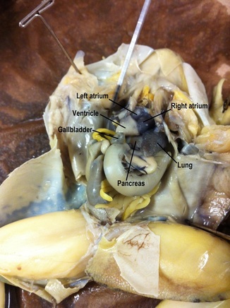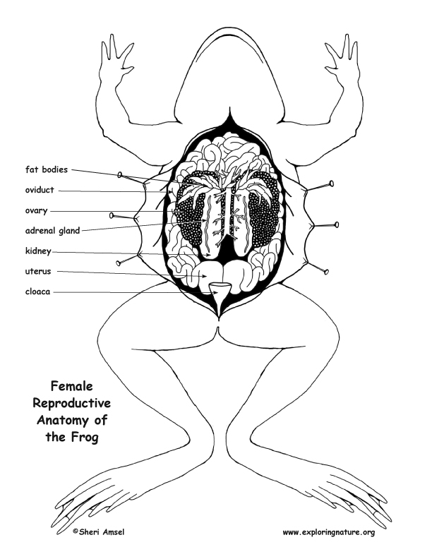
Follow the grasshopper through the frog, placing the organs that are used for digestion in order. Created from high-quality, real images, this excusive dissection design. In layer four, students examined the kidneys and spleen. Frog Dissection Diagrams & Questions Name: Teacher: Hour: 2 The frogs that we are going to observe have never lived in the wild. The hind limbs are in the rear of the frog and are much longer and more muscular than the forelimbs located anteriorly. Conduct a detailed frog dissection using ultra-realistic 2D paper illustrations. In layer three, students identified the lungs and the pancreas. Frog Dissection Diagrams Flashcards Quizlet Science Biology Anatomy Frog Dissection Diagrams 4. Web this is a printable worksheet called frog dissection labeling and was based on a quiz created by member eleni yargo. Cut along the indentations that separate the thoracic portion of the carapace into three regions. Lyons pointed out that the heart and the liver hid some of the organs that students would identify in layer two. Web the frog’s reproductive and excretory system is combined into one system called the urogenital system. Using one hand to hold the crayfish dorsal side up in the dissecting tray, use scissors to carefully cut through the back of the carapace along dissection cut line 1, as shown in the diagram below. In layer two, students identified the gallbladder, stomach, and the small intestine. In layer one, they identified the liver, the heart and the three chambers of the heart. In dissecting the internal anatomy, students looked at the organs in four different layers. In examining the external anatomy students identified the upper and lower jaw, the tongue, the maxillary teeth, and the glottis. In addition to the dissection, students identified all of the parts of the frog, and labeled the parts on a printed diagram. Internal Anatomy of the Frog Labeling (BW)5. Internal Anatomy of the Frog Diagram (BW)4. Internal Anatomy of the Frog Labeling (Color)3.

Internal Anatomy of the Frog Diagram (Color)2.

Now select Show male at the bottom left to switch to the male frog. This includes 18-pages of Frog Internal Anatomy Diagrams, Labeling Pages, and Activities in color and black and white.1. With the rotate button selected, click and drag on the frog to rotate it. Students examined the external anatomy, mouth, internal anatomy, muscle and bone, and internal body systems. In the Frog Dissection Gizmo, you will complete a virtual dissection of a female and male frog. Lyons’ as he demonstrated the frog dissection using his document camera. Students took turns assisting and dissecting as they were directed by Mr. Lyons’ seventh grade science classes worked in pairs as they took part in frog dissections on Wednesday December 18th and Thursday December 19th. Lyons’ classroom on Wednesday, he was handing out frogs to student groups for the dissection lab.
#Frog dissection diagrams full#
Some individuals do not enjoy performing dissections of full organisms, but instead. It is important to determine which type of dissection is best for your student or child.

color the gall bladder green and the bile duct (3b) a darker green. Frog dissections are a great way to learn about the human body, as frogs have many organs and tissues similar to those of humans. Tucked under the liver is the gall bladder, which stores bile that is produced by the liver.The liver has several jobs related to digestion and detoxification. Periodically, your instructor may pause to show you illustrations, diagrams or videos of. Color the left anterior lobe (b) medium brown, and the left posterior (c) lobe dark brown. In the lab, you will be spending a few days, dissecting the frog. The largest organ is the liver, and it consists of multiple lobes.Leading from the mouth is a tube that connects to the stomach.When the abdominal cavity of the frog is opened, many organs of the digestive and urogenital systems can be observed.Īs you read the descriptions of the organs below, color them on the diagram.


 0 kommentar(er)
